At Southern Eye Centers, we believe great care begins with understanding your needs. Whether you visit us in Baton Rouge or Plaquemine, our Louisiana optometrists take time to evaluate your vision, protect your long-term eye health, and recommend personalized solutions. Families across the region trust our team because we combine advanced diagnostics with a welcoming, patient-first experience you can count on.
Quality Eye Care in Baton Rouge & Plaquemine, LA

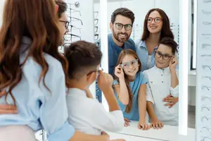
Trusted Eye Doctors for Capital Region Families
Our practice offers vision care for adults, seniors, and children as young as three, making it easy to manage everything from routine eye exams to ongoing medical needs in one place.
Your Plaquemine optometrist or Baton Rouge eye doctor will explain your results, answer your questions, and guide you toward solutions that support your eyesight and overall wellness.
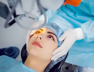
Emergency Eye Care
Eye problems don’t always happen at convenient times. Southern Eye Centers sets aside same-day appointments and provides after-hours support for urgent concerns. Call us if you experience sudden vision changes, flashes of light, new floaters, eye pain, redness, chemical exposure, or an eye injury.
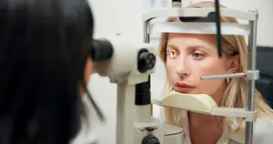
Diagnostic Technology That Supports Better Outcomes
Southern Eye Centers invests in modern equipment that helps us detect changes earlier, measure vision more accurately, and monitor eye conditions with exceptional detail.
Tools such as retinal imaging, OCT, and corneal topography enable our Baton Rouge eye doctors to accurately evaluate your eye health and create treatment plans tailored to your unique needs.
Whether you’re receiving advanced dry eye care in Baton Rouge or routine monitoring in Plaquemine, our technology helps ensure your care is both thorough and dependable.

Optometry Services at Southern Eye Centers
Routine evaluations that protect your vision and support lifelong eye health.

Protect your vision with a thorough diabetic eye exam for long-term eye health.

Protect your vision with glaucoma exams that detect early optic nerve changes.


Kid-friendly care for children ages 3 and up, focused on clear sight and early detection.

Specialized care using modern imaging and advanced contact lens expertise.